Select a category
Advertisement

Diabetic Foot Examination (Surgery Rotations)
4 years ago
Advertisement
Foot Examination
1. Define Diabetic foot
A constellation of Physical findings and Medical complications in the foot, arising as a consequence of Several conditions induced by diabetes such as (VIP WIN)
* Venous Stasis
* Immobility of the chronically ill diabetic (Cant care or inspect and increased pressure on points)
* Impaired blood supply due to concurrent PVD
Impaired Wound healing and secondary infection due to relative Immunosuppression
* Impaired sensation due to diabetic Neuropathy
Diabetic Foot is a common cause of hospitalization and leading cause of amputation in US
2. Inspection (KACHA THIN DOCS)
* Legs exposed above Knees
* Look for Prior Amputation
* Cleanliness: Leg Hygiene (Inadequate hygiene is a risk factor for infections)
* Hair pattern (Difference btw legs, Abrupt change in hair pattern as you go down the feet)
Ø May indicate Peripheral vascular disease
* Around the Foot:
Ø Plantar surface
Ø Dorsal surface
Ø Medial surface (Medial Malleolus, Ball)
Ø Lateral surface
* Tips of toes
* Heel
* In between toes/Interdigits
Ø Commonly see Superficial Fungi Infection)
* Nails
* Joint Deformities
Ø Metatarsal subluxation
Ø Hammer toes
Ø Halux adductor valgus or Rigidus
Ø Abnormally high Arch etc.
* Osteomyelitis:
Ø Is Bone visible within the ulceration? OR
Ø Can bone be Reached with a probe (Osteomyelitis is most likely)
* Calluses, Corns
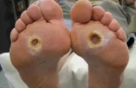
* Sores/Ulcerations;:
Ø May be due to
· Trauma from Foot wear
· Arterial insufficiency
· Venous stasis
· Neuropathy
Ø Don’t forget to Describe and Measure the ulcers
Summary: Inspection of an Ulcer (What to look out for)
KACHA THIN DOCS
Knee (Expose foot ulcer to above knee)
Amputation (Look for prior amputation)
Cleanliness
Hair pattern/Growth
Around the foot
Tips of toes
Heels of foot
Inter-digits area
Nails
Deformities
Osteomyelitis
Calluses/Corns
Sores/Ulcers
3. Detecting Neuropathy (UVA/B MTG SCAN)
n Look for Ulcers on the Plantar surface of the feet (Indicate neuropathic disease)
n Prominent Veins (Often in dorsum and Lower calf) – due to arteriovenous shunting in diabetic neuropathy and autonomic dysfnx = widely dilated vascular bed. (Pt should stand)
n Abnormal sensations; Numbness, Prickling, Tingling, burning, throbbing or shooting pain, Feet is warm,
loss of Balance and coordination.
n Bounding pulses
n Reduced Sweating; suspect if
Ø Dry skin (from decreased sweating),
Ø Fissures (from decreased sweating);
Ø Dec. sweating due to autonomic dysfnx
n Claw toes (from wasting of small muscles)
n Arch high (from wasting), Arch low (From Charcot foot)
n Test for Neuropathic sensation of feet (P-TPTP) and Ankle reflex
Ø Touch (wool)
Ø Pain (Sharp)
Ø Temperature (Cool and warm surfaces – can use Steel)
Ø Pressure (Blunt edge)
Ø Proprioception/Balancce: pt should know direction of passive toe motion without looking.
n Test for Ankle reflex
n Use 10gram mono filament to test for loss of sensation
n Apply tuning fork over bony prominence to test for loss of vibration
n Assess gait for smoothness and symmetry
4. Detection of Peripheral Arterial Disease (CAT PURA)
n Inspect the foot
Ø Look for Color changes
Ø Arteries: Palpate Dorsalis pedis artery (Lateral to the tendon of Extensor hallucis longus/Medial to the tendon of extensor digitorum longus tendon)
Ø Look for Ulcer location (Ulcer on foot side indicate Neuro-ischemic disease)
n Palpation (Temperature difference, Dorsalis pedis artery and Post. Tibial Artery) and Hair pattern
Ø Compare temperature on both feet and other normal areas
Ø Palpate posterior Tibial artery (Mid way btw Medial Malleolus and Achilles tendon) with foot passively dorsiflexed.
n Assess capillary Refill (With Nail of great toe); Of little value in assessing for PVD
n Assess more Proximal pulses (Popliteal pulse: Midline within popliteal fossa, behind knee, best felt with hands wrapped around the knee while knee is relaxed in moderate flexion) if distal ones are abnormal
n Describing pulses: Absent, Present but Diminished, Normal, Bounding. (BAND)
n If suspect any PVD, Measure systolic ankle pressure (Sphygomanometer and Hand held doppler)
Ø Note the highest and Lowest pressure
n Calculate the Ankle Brachial Index (ABI = Highest pressure at Ankle/Highest Systolic pressure of Brachial artery)
n Defective feet perfusion dx if ABI < 0.9
Summary: Checking for Peripheral Arterial Disease
CAT PURA
Color changes
Arteries palpated
Temperature difference
Proximal pulses assessment
Ulcer location(s)
Refill (Capillary refill)
Ankle Brachial index calculation
5. Foot deformities (CHHAM)
Can Cause increase in foot pressure
n Inspection
Ø High Arched foot (Muscle wasting)
Ø Metatarsal head prominence (Metatarsalgia/ Football overuse injury)
Ø Halux adductor Valgus or Bunion (A hallux valgus deformity, commonly called a bunion, is when there is medial deviation of the first metatarsal and lateral deviation of the great toe (hallux). The condition can lead to painful motion of the joint and shoe wear difficulty). Bunions are not diabetic lesion but can complicate the tx of DM foot
Ø Halux Rigidus (Hallux rigidus is a disorder of the joint located at the base of the big toe.
Advertisement
Ø Hammer toes (Clawing)
Ø Neuro osteoarthropathy (Charcot foot)
Charcot foot is a condition causing weakening of the bones in the foot that can occur in people who have significant nerve damage (neuropathy). The bones are weakened enough to fracture, and with continued walking, the foot eventually changes shape (Repetitive trauma due to insensitive joint). Often causes ROCA bottom deformity (fallen/everted arch – worse case), and most likely leads to ulcer formation on deformed area.
· Early signs: Edema, Warmth, Erythema surrounding affected joints (Inflammatory)
· Late: Overt joint deformity, dislocations, pathologic fractures, overlying skin ulcerations
· Not specific for diabetes also seen in other etiologies of PVD: Alcoholism (B9 def), HIV, Syphilles (Tabes dorsalis), B12 def (SCD).
· Suspect acute Charcot foot in any patient with neuropathy if they present with Hot, Swollen and Painful foot.
Summary: Common Foot deformities in Diabetic foot
CHHAM
Charcot foot
Halux rigidus and Halux adductor valgus
Hammer toes
Arched (Abnormally high) foot
Metatarsalgia
6. Non-Ulcerative Pathology (BMC)
Ø Bullae (A bulla is a fluid-filled sac or lesion that appears when fluid is trapped under a thin layer of your skin.
Advertisement
Ø Maceration (Maceration occurs when skin is in contact with moisture for too long. Macerated skin looks lighter in color and wrinkly. It may feel soft, wet, or soggy to the touch. Skin maceration is often associated with improper wound care.)
Ø Callous (A callus is an area of thickened skin that forms as a response to repeated friction, pressure, or other irritation.
Advertisement
· Hemorrhages within callus is an important precursor of ulceration
· Corns may be wrongly referred to as calluses, but they are different.
a. Corns are smaller than calluses
b. They are usually surrounded by an inflamed skin
c. They seldom develop on weight bearing points on the feet (There may be found on top the foot, sides or in between the toes)
7. Nail abnormalities (DIT)
Ø Deformed nail
Ø Ingrown nail
Ø Thick nail
8. Ulcers - Locations (DIPS)
Ø Plantar surfaces ulcers
Often due to neuropathy (Patient doesn’t shift weight)
Ø Sides/Medial and Foot side ulcers (Around the Maleolus, In step, ball of the foot)
Often due to Neuroischaemic disease
Ulcers can appear on other sites
Ø Interdigital and Dorsal surfaces ulcers
Can be caused by tight, ill fitting, shoes in both neuropathic and Neuro-ischaemic feet
Summary: Common locations of ulcers in Diabetic Foot
DIPS (All sides of the foot)
Dorsal surfaces
Interdigital surfaces
Plantar surfaces
Sides of the foot
9. Detecting infection (5 Signs of Inflammation; + Discharge, Smell)
n Infection requires diagnosis and therapeutic measures
n Signs of infection may be lacking in people with diabetics
n Inspect and palpate for foot changes, warmth, swelling, discharge, Bad smell and pain. Maceration due to fungi infection, foot wear.
10. Instruct patient about foot care before they leave the clinic
11. Frequency of follow up visits (Depends upon Risk Categorization)
n No sensory neuropathy
Ø Visit/Follow up once a year
n Presence of Sensory neuropathy
Ø Visit/Follow up every 6 months
n Presence of Sensory Neuropathy + Peripheral Vascular Disease + Foot deformity
Ø Visit/Follow up every 3 months
n History of Previous Ulcer + Amputation
Ø Visit/Follow up is done monthly
12. Management of non-ulcerative lesions
n Cutting and Trimming of nails
n Callus
Ø Pay attention to callus
Ø Ulcer is imminent if neglected or treated inappropriately
Ø Stretch the tissue and ensure cutting/even removal of callus (layer by layer)
n Pressure assessment with custom made soles
n Bullae
Ø Drain tense ones by cutting across the roof of the bullae
Ø Evacuate the content and cut off the overlying skin
Ø Inspect the base
13. Management of Ulcer (DEMA)
n Excision/Remove calluses surrounding the ulcer
n Slough/Cut away all non-viable tissue
n Debride from middle outwards
n Probe the base of the ulcer (If probe reaches bones, suspect osteomyelitis)
n Clean ulcer with normal saline
n Take a swab of wound sample and send for culture
n Measure the ulcer, two diagonal measurements
n If Ulcer is irregular, trace the outline (With transparent and white paper, discard the transparent paper)
n Bleeding after ulcer debridement (Tx with Pressure, Limb rest and Calcium arginate)
n Calcium arginate (Alginate dressings are dry when initially placed on an open wound and become larger and more gel-like as they draw in fluids. ... Alginate dressings can also help wounds that are bleeding. The calcium in these dressings helps stabilize blood flow, which slows bleeding)
n Cover with light bandage (Don’t wrap too tightly, and don’t encircle the toe)
n Review and repeat at regular intervals
Summary: Management of Ulcers
DEMA
Debridement and Sloughing
Excision of calluses
Measurement
Arginate (Calcium) dressing preferable.
14. Offloading (Relief of mechanical pressure)
n Total contact caste
n Removable cast walker
n Cast shoes
n Therapeutic footwear
15. Predictors of amputations (HAND)
n High Hb A1c (N: 4 – 5.6%, DM risk: 5.7 – 6.4%, High: > 6.5 - > 7)
n Amputation (Presence of a previous amputation)
n Neuropathy and Impaired arterial supply
n Deformities/Ulcers present
16. Three major components of DM foot exam
n Inspections
n Assessment of Vascular Arterial supply
n Assessment of Neurologic fnx
17. The following signs don’t necessarily indicate infection of an ulcer but should prompt evaluation for infection
n Erythema
n Tenderness
n Purulent drainage
n Smell
18. Ulcer
n Acute ulcer
Ø Ulcer present for less than six weeks (< 6 weeks)
n Chronic ulcer
Ø Ulcer present for more than six weeks (> 6 weeks)
My Summary of Diabetic Foot Exam
· Define: A constellation of Physical findings and Medical complications due to one or several conditions induced by diabetes. These Conditions include VIP WIN
· Predictors of Amputation:
Ø HAND
· Indications of Amputation (4):
Ø Dead
Ø Deadly
Ø Dead Loss
Ø Damn nuisance
May also remember with GOMBE LIONS
· Complications of Amputation include:
Ø Local and surgical complications
Local complications of Amputation: DISH WOOPS
· Components of DF exam
1. Inspection
2. Neuropathy exam
3. Vascular exam
4. Nail exam
5. Deformities exam/note
6. Non-ulcerative lesions exam
7. Ulcer exam
8. Mgt & Prevention
.Disclaimer If this post is your copyrighted property, please message this user or email us your request at [email protected] with a link to this post
Advertisement
 Peter
Peter